Force Myographic Interface for Ankle Rehabilitation
Tiffany Morris, MS. and William Craelius, PhD.
Department of Biomedical Engineering – Rutgers The State University of New Jersey
Piscataway, NJ 08854
ABSTRACT
Neuromuscular rehabilitation regimens can be demanding and tedious for users. Programs that link physical exercise with game playing or simulated activities within virtual reality environments attract many users. Herein we describe a non-invasive interface to a computer mouse emulator using force myography (FMG). The interface consists of a pressure-cuff around the calf, and can be used to control the cursor for game playing and computer utility.
KEYWORDS
Neuromuscular, force myography, muscle, biofeedback, ankle
BACKGROUND
Neuromuscular rehabilitation is a necessary for many populations including those with stroke and cerebral palsy. To restore mobility therapists have used several approaches including muscle strengthening, biofeedback, anti-spasticity treatments, orthotics, and robotic devices. Essential to promoting effective motor learning during these therapies is proper sensory feedback; however, neuromuscular deficits usually limit such feedback. Alternate channels of feedback are thus desirable, so that appropriate muscle actions can be rewarded and inappropriate ones discouraged. The usual mode of muscle registration for biofeedback is the EMG, which has been successful (1). These biofeedback protocols have been enriched using virtual reality environments and computer game playing, in order to encourage users to perform the arduous physical therapies. To extend these promising, but expensive, strategies to the widest target audience new tools are required.
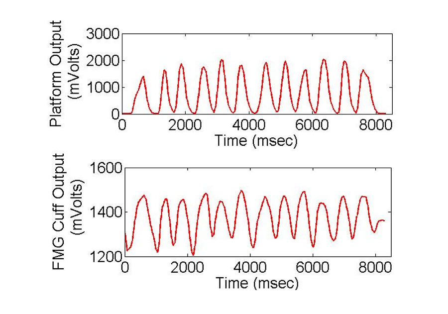 Figure 1: FMG correlation to ankle flexion. (Click for larger view)
 Force myography (FMG), the measure of external muscle pressure, is a simple and reliable method of detecting and providing feedback of muscle activation(2). During concentric and eccentric contractions of muscles close to the surface, changes in muscle diameter are visible by eye and can be detected using force transducers such as, force sensing resistors (FSR). Compared to EMG, FMG is simple to apply and process, and can register activation of large muscle groups adequately for real-time biofeedback .To date FMG has been implemented by only a few groups. Amft et al. created an interface capable of detecting grasp activity by securing the FSRs to the skin with tape, over two large muscle bellies(3) Yungher, et al. used an array of FSRs mounted on a cuff to detect pressures in the leg of a single female subject as she walked on a treadmill(4). Both groups found that the FMG signals corresponded to the EMG signals. Additionally, it was previously established that there is a good correlation to the FMG signal of the calf muscle and ankle rotation (rho=.81)(5).
RESEARCH QUESTION
Our strategy herein is to combine a FMG biofeedback interface with a mouse adapter, enabling patients to play preexisting mouse controlled games using wrist, elbow, or ankle flexion motions. In this project we seek to determine whether our current FMG interface is suitable for mouse cursor control. We hypothesize that the current FMG interface is suitable for mouse cursor control, and together with the mouse emulator will provide an engaging environment for physical rehabilitation.
METHOD
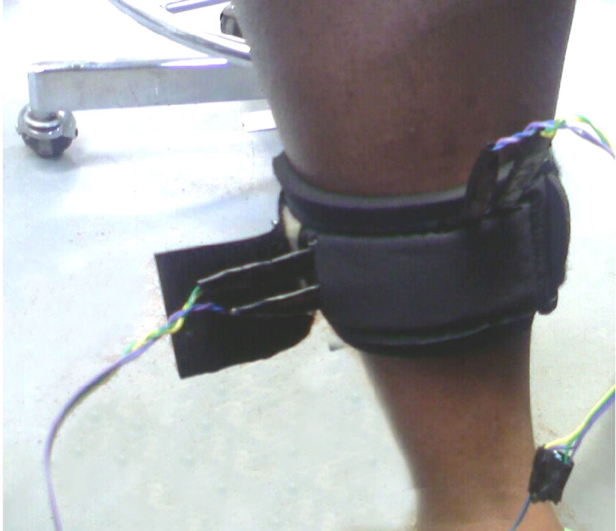 Figure 2: FMG cuff. (Click for larger view)
 Six non-impaired subjects were recruited to test the device. As a pilot one stroke subject tested the device. Six suitable mouse controlled internet games were downloaded from the internet for testing. A suitable game was said to be any game that involved only two directions of movement. Each subject was asked to complete a questionnaire, designed to collect basic subject demographic data such as activity level and the amount of time spent playing computer games. Written informed consent was obtained from all participants. This study was approved by the Rutgers University IRB.
The FMG cuff hardware is comprised of four force sensitive resistors placed within a standard wrist support band. The FSRs are connected to a silicon laboratories microcontroller board, which is used to power the force sensitive resistors, and to convert the analog voltage signals to a mouse input. A LabVIEW program was created to run games and record the FMG signals.
Each subject used two FMG cuffs, one for each calf. The mouse cursor position was proportional to the FMG signals received from each calf muscle. When the ankles were in a neutral position (feet flat on the floor), the mouse cursor remained in the center of the screen. As the left ankle was dorsiflexed the cursor moved to the left of the screen in proportion to the flexion, and as the right ankle was dorsiflexed the cursor move to the right of the screen in proportion to the dorsiflexion. Before playing the games patients familiarized themselves with the device by controlling a cursor on the computer screen. Subjects then selected a game of their choice and either ran a practice session in which they familiarized themselves with the game, or played the game while ankle flexions were recorded.
RESULTS
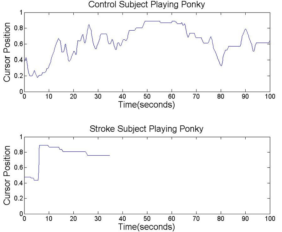 Figure 3: Sample Ankle Movement Recordings. (Click for larger view)
 All healthy subjects successfully controlled the mouse cursor using ankle flexion. The stroke subject had difficulty controlling the cursor using ankle flexions without a mechanical support. Figure 1 shows the FMG signals of a stroke and healthy subject while playing on round of the Ponky car racing game. The ordinate shows the normalized mouse cursor position, with 0 indicating the left most cursor position and 1 indicating the right most cursor position. The stroke subject made fewer flexions and had a shorter round duration (36 seconds) than the healthy subject (100 seconds). Also, the stroke subject made only one flexion with the affected leg and tended to use the un-affected leg for most of the cursor control.
The four most popular games, Trickmaster, Ice racer, Watch out and Ponky, were studied in more detail. The relationship between the length of time the games were played and the age of the subject was studied. The games were divided into two categories, sport and juvenile. Sport games were defined to be games that mimicked playing a sport, such as Trickmaster (skateboarding), and Iceracer ( bob sledding). Juvenile games were defined to be games that looked more like cartoons, and did not attempt to mimic a real life situation, such as Ponky (a car racing game), and Watchout (a game in which the player dodged falling boulders). Although everyone played both types of games, four out of six of the subjects spent more time playing the juvenile games.
DISCUSSION
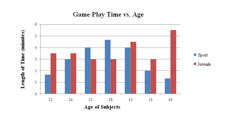 Figure 4: Relationship Between Game Play Time and Age (Click for larger view)
 We demonstrated that healthy subjects can control a mouse cursor with ankle motions registered by FMG. Using FMG signals to control a mouse cursor made it easy for us to include games that satisfied people of different ages. Ankle flexions proved to be very difficult for the stoke subject. Flexing the ankle against gravity is a difficult task for many stroke patients, therefore a mechanical assist may be necessary when working with ankle flexions. However, it may be possible to use wrist or elbow flexions without an assist. When given the option to use the both legs, it is likely that the patients will rely more on the unaffected leg for cursor control, as seen with our subject. Therefore, we have begun designing a system that can control both directions with one limb. We believe that making a system in which relaxing a muscle, and activating a muscle controls the two directions of the mouse cursor may be more effective.
REFERENCES
- Moreland, J.D., M.A. Thomson, and A.R. Fuoco, “Electromyographic biofeedback to improve lower extremity function after stroke: a meta-analysis”, Arch Phys Med Rehabil, 1998. 79(2): p. 134-40.
- Phillips, S.L. and W. Craelius, “Residual kinetic imaging: a versatile interface for prosthetic control ”, Robotica, 2005. 23: p. 277-282.
- O. Amft, H. Junker, P. Lukowicz, G. Troster, and C. Schuster, “Sensing muscle activities with body-worn sensors,” in Proc., 2006.
- D. Yungher, M. Wininger, A. Threlkeld, J. Barr, and W. Craelius, “Force myography analysis of leg muscle activity,” in Proc. American Society of Mechanical Engineers Summer Bioengineering Conference, 2006.
- T. Morris, S. Escaldi, C. Glass, N.A. Newby, and W. Craelius, “Quantifying Ankle Motion Using The Ankle-Foot Interface (AFI),” in Proc. 12th World Congress of The International Society for Prosthetics and Orthotics, 2007.
ACKNOWLEDGMENTS
This study was funded by the National Institute on Disability and Rehabilitation Research grant # H133E030011-06 (RERC) granted to the New Jersey Institute of Technology. We would like to thank the physical therapists at Children’s Specialized Hospital for their input, and Nam Hun Kim, Ph.D.
Author Contact Information:
Tiffany Morris, MS, Rutgers University, 599 Taylor Rd., Piscataway, NJ, 08823, Office Phone (732) 445-2369 EMAIL: tiffm@eden.rutgers.edu